Le femur, forming on its own the skeleton of the thigh, is the longest bone in the human body. It is also the largest and most resistant, because it has to withstand very high pressures (up to 280 kilograms/cm² during a jump).
Although strong, the femur can "break". The broken bonesdufemur are generally the result of violent trauma, most often occurring in the context of a traffic or public highway accident.
They can, however, occur as a result of low-energy trauma when there is ground for fragilitybone, especially in the elderly or in certain pathologies such as osteoporosis.
Go directly to the section that interests you
femur anatomy
The femur is a large, elongated bone. We have two femurs, each one consists of three parts:
Proximal epiphysis: it is the upper end of the femur. It is made up of a neck (neck of the femur) and a head which articulates with the acetabular cavity (articular surface of the pelvic bone: the coxal bone) forming the coxo-femoral joint (the hip ).
Diaphysis: it is the central part of the femur located between the two epiphyses.
Distal epiphysis: lower end of the femur. It articulates with the tibia and the patella forming the femoro-tibial and femoro-patellar joints at the level of the knee.
What is a femur fracture?
It is any fracture whose line sits at the level of the femur. Depending on the location of this feature, there are three categories of femoral fractures:
Fractures of the upper end of the femur
Fractures of the upper extremity of the femur represent the most common trauma emergency in the elderly. They are classified into two main varieties:
Femoral neck fractures: these are also called cervical fractures of the femur. The fracture line sits at the level of the neck of the femur, that is to say the bony portion which connects the head of the femur to the femoral diaphysis.
Trochanteric fractures: they sit in the region between the femoral diaphysis and the neck-head complex of the femur. They can concern the lesser trochanter, the greater trochanter (bony projections) or any other area of the trochanteric massif.
Fractures of the femoral shaft
Fractures of the femoral shaft frequently fall within the scope of a polytrauma (several injuries including at least one life-threatening) due to the generally violent nature of the accident.
Fractures of the femoral shaft can be classified according to the location of the fracture line in :
Diaphyseal fractures of the upper third of the femur.
Diaphyseal fractures of the middle third of the femur.
Diaphyseal fractures of the lower third of the femur.
They can also be classified according to fracture line type in :
Simple fracture: a single fracture line separating the femur into two bone fragments. It can be transverse, oblique or spiroid (in torsion).
Complex fracture: there are at least three bone fragments.
Bifocal fracture: there are two fracture sites (the bone breaks in two different places).
Comminuted fracture: it is a complex fracture that usually results from a violent trauma fragmenting the bone into several small pieces.
Fractures of the lower end of the femur
They are generally the prerogative of the young subject, following violent traumas such as traffic or public highway accidents.
This type of fracture can also be seen in elderly subjects for less violent traumas such as ordinary falls or moderately violent shocks, even light ones.
What are the causes of femur fracture?
Femur fractures, as mentioned above, usually occur in young people after a traumaviolent. Thus, they can be seen in the following circumstances:
There are also what are called the "pathological fractures". They occur for minimal trauma, even spontaneously (without any trauma). They are generally observed in a context of bone fragility, particularly in the context of osteoporosis or secondary to taking certain medications (long-term corticosteroids, for example).
The causes of femur fractures can be classified into two main categories: direct causes and indirect causes.
Direct causes
In this category, fractures occur directly at the point of impact. There are mainly two situations:
The crash: trauma during which the thigh is caught in a vice between two bodies. For example, a car wheel crush, the first body being the wheel which represents the injuring agent, and the second being the ground which represents a support.
Direct shock: the fracture occurs directly at the point of impact.
Indirect causes
Here the fractures occur distance from point of impact. Let's take as an example a fall from a height of about ten meters with landing in a standing position. The point of impact is therefore represented by the two feet. The energy of the impact will be transmitted throughout the lower limbs, which can cause fractures in any part: ankle, leg, knee, thigh...
What are the symptoms of a femoral fracture?
The clinical elements that point to a fracture of the femur are:
Evocative context: the circumstances of the accident (fall, road accident, etc.).
Cracking: a sensation of cracking in the thigh at the time of the shock can be described by the patient.
Mechanism of trauma: direct or indirect.
Total functional impotence of the lower limb: impossibility to lean on the injured limb, let alone walk.
Thigh pain at the fracture site: diffuse pain in the groin in case of fracture of the femoral or trochanteric neck…
Thigh deformity: is seen especially during so-called "displaced" fractures, that is to say with loss of alignment of the bone fragments.
Rotation of the lower limb: the different muscles pull the bone fragments towards them. In the event of a femur fracture, this results in external rotation of the injured leg (by the action of the gluteal muscles and lateral hip rotators).
Shortening of the lower limb: by overlapping of the two femoral bone fragments.
Complications: sometimes, femur fractures are complicated by:
Cutaneous opening (open fracture).
Vascular lesion (damage to a blood vessel).
Nerve damage (damage to a nerve).
Associated fractures (pelvis, upper limbs, etc.).
Associated lesion involving the vital prognosis (mainly head trauma, chest, abdomen, etc. This is called polytrauma).
How is a femoral fracture diagnosed?
The diagnosis of a femur fracture is usually easy to make. The elements of the interrogation (notion of trauma, pain, total functional impotence of the lower limb, etc.) supplemented by the data from the physical examination of the patient (deformation of the lower limb, pain caused by pressure, etc.) quickly lead to the diagnosis. .
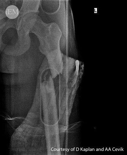
In order to confirm the latter, the doctor asks for a standard x-ray oflathigh front and profile. This will highlight the fracture site(s) in the femur. It will also make it possible to classify the fracture (several classifications exist in order to standardize the management of the various fractures throughout the world according to the data of scientific studies).
In current practice, other X-ray images are systematically requested, in particular an X-ray of the jointstheir et under-underlying (knee and hip) as well as an x-ray of the basin from the front (to check the integrity of the pelvis and the hip and knee joints: search for associated lesions).
Sometimes, X-rays do not reveal any bone abnormality despite the patient's painful complaints and the context strongly suggestive of a femoral fracture. In this case, it is possible to make a scanner with bony window to clarify the diagnosis.
Management of a femur fracture
It's the orthopedic doctor which supports femur fractures. To choose the most suitable treatment for the patient, this specialist takes into account many parameters:
The seat of the fracture (distal or proximal end, diaphysis, neck, trochanteric region, etc.).
The type of fracture (simple, complex, displaced or not displaced, open or closed, etc.).
Possible lesions associated with the femoral fracture.
The patient's age.
The quality of the patient's bones.
General state of health.
orthopedic treatment
In children, we generally recommend a traitementorthopedic in the event of a simple fracture of the femoral diaphysis. This is based on the reduction of the fracture followed by immobilization of the lower limb in extension in order to allow its consolidation.
Reduction of the fracture can be achieved by various methods. Sometimes, a simple traction of the lower limb puts the bone back in place. In the event of failure, the patient is taken to the operating room, put to sleep (general anesthesia or deep sedation) then an attempt is made to align the femur using maneuversexternal with regular radioscopic verification (thanks to a device that allows the production of instant x-rays displayed on a screen).
After obtaining a reduction deemed satisfactory by the orthopedist, the lower limb is immobilized with a plasterpelvis-pedal. The latter extends from the hip to the foot in order to allow the immobilization of the hip and knee joints.
In young children, it is necessary to count 6 to 8 weeks of immobilization for a stable, non-displaced femur fracture.
Surgical treatment
In adults, femur fractures are treated surgically. The treatment can use various methods:
Intramedullary nailing: it consists of placing a nail at the level of the medullary canal of the femur in order to maintain the bone fragments in the correct position.
Osteosynthesis by screwed plates: it is widely used in fractures of the upper extremity of the femur. It consists of realigning and fixing several bone fragments with a metal plate and screws.
Arthroplasty: consists of replacing the femoral head and the neck with a prosthesis. It is indicated in certain fractures of the upper end of the femur.
Re-education
Rehabilitation sessions by physiotherapy are always necessary after surgical treatment of a fractured femur. They must be started as soon as possible in order to prevent stiffness joints (particularly at the level of the hip and the knee) and allow the patient to regain strengthmuscular and normal mobility.
Depending on the type of intervention undergone, the resumption of weight bearing can be done in a few days to several weeks. To prevent the risk of thromboembolic complications (venous thrombosis, pulmonary embolism, etc.) postoperatively, the patient is placed under traitementanticoagulant during the immobilization period.
The healing time of a femur fracture in adults depends on several factors. She is from three months on average.
References
[1] A. Lambotte, “Operative surgery for fractures”, Masson, 1913.
[2] A. Vannineuse and C. Fontaine, “Fractures of the proximal end of the femur” Springer Science & Business Media, 2000.
[3] JC Bel and L. Fischer, “History of the treatment of femoral neck fractures”, Rheumatol. Prat., p. 33-36, 2009.
[4] Father Simon et al, "Fractures of the neck of the femur after 50 years", Rev. Chir. Orthopedic Restorative Appar. Word., flight. 94, no 6, p. 108-132, 2008.
[5] G. Lamraski, D. Toussaint, and J. Bremen, “Surgical treatment of fractures of the lower end of the femur by extramedullary osteosynthesis”, Acta Orthop. belg., flight. 67, no 1, p. 32-41, 2001.
My name is Katia, I am specialized web editor in writing medical articles. Being passionate about medicine and writing, I set myself the goal of making medical information accessible to as many people as possible, through the popularization of even more complex scientific concepts.
My name is Katia, I'm 27 years old and I'm a web writer specializing in writing medical articles. Being passionate about medicine and writing, I have set myself the goal of making medical information accessible to as many people as possible, thanks to the popularization of even more complex scientific notions.

")


")
")
")