Article reviewed and approved by Dr. Ibtissama Boukas, physician specializing in family medicine
La Legg-Perthes-Calvé disease ou primary osteochondritis of the hip (OPH) is a pathology of the joint of the hip which affects children from 2 to 12 years old. It is found mainly in the age group of 5 to 6 years. Boys are found to be more affected than girls.
If you have a child who suffers from hip without any previous trauma, think about the Legg-Perthes-Calvé disease. This article will take care of enlightening you a little more about this disease. The causes, symptoms, diagnostic means and means of treatment will also be discussed.
Go directly to the section that interests you
Definition and anatomy
Definition
La Legg-Perthes-Calvé disease is the term used to define death (necrosis) of the hip bones in children. Bone death therefore concerns the femoral head as inhip osteonecrosis in adults. This necrosis occurs following a low blood supply in the growth cartilage (growth cartilage) of the head of the femur. What is growth plate?
Anatomy
Before talking about the growth plate, which is the anatomical structure involved in the Legg-Perthes-Calvé disease, let's do a little reminder on the anatomy of the hip. A more complete anatomical description of the adult hip is made in the article onhip osteonecrosis.
The hip is the joint that connects the trunk to each lower limb through the union of the pelvis and the thigh. This is the coxo-femoral joint (coxal bone for the pelvic bone and femur for the thigh bone). It is a joint that plays a very important role in the statics and dynamics of the body. It must be stable while carrying the load of the trunk when walking and when standing.
There are two hip joints (left and right). The hip joint is made up of many anatomical elements. First of all there are the two articular surfaces of the ends of the two hip bones (coxal bone and femur).
The acetabulum or acetabulum: it is the hemispherical cavity of the external face of the coxal bone. It articulates with the head of the femur.
The head of the femur: It has a spherical shape and fits perfectly with the acetabulum.
Then there are the other parts of the femur which are:
The femoral neck: it is located between the femoral head and the greater trochanter.
The greater trochanter: It is a voluminous bony projection in the extension of the body of the femur called diaphysis.
The lesser trochanter: is a small conical shaped bony prominence below and posterior to the femoral neck.
The growth plate
Growth cartilage or growth cartilage is a cartilage that only exists in growing children. It is located between the epiphysis (head) and the diaphysis (body) of long bones such as the femur. It is this anatomical element that allows the development in length of the bone when the child grows in size.
Growth cartilage develops under the influence of growth hormones and induces bone formation at the expense of cartilage. In addition to the ability of our brain (hypophysis) to synthesize growth hormone, genetic heritage, nutritional resources, diseases, stress, are other factors that affect the growth of our bones.
This process of long bone development continues until adulthood when the growth plate has completely transformed into bone and is fused together. Adults therefore no longer have growth plate.
Causes of Legg-Perthes-Calvé disease
The causes of Legg-Perthes-Calvé disease are poorly known. All that is known of the mechanism is that there is destruction of the head of the femur in one or more places due to lack of blood supply. Although real causes have not been found, a few factors could favor this state of destruction of the femoral head.
Delayed maturation of the frame in children
Delayed and disproportionate growth of the child
A small child
Low birth weight
A state of malnutrition due to social and economic deprivation
A trauma
Birth defects
Genetic abnormalities (mutations of the gene COL2A1)
Coagulation abnormalities such as thrombophilia (tendency to thrombose) or hypofibrinolysis
Venous thrombosis (blood clot in a blocked vein).
Symptoms of the disease
The main symptoms at the start of the Legg-Perthes-Calvé disease are :
La lameness
A pain in the groin, thigh or knee
A limitation of movements of the hip.
At a more advanced stage of the disease, it may appear:
A length inequality between the two lower limbs
A muscle atrophy (decrease in the volume of the muscles) at the level of the hip concerned.
The other signs of the advanced stage are more visible in the paraclinical (standard X-ray images). It is :
Deformity of the femoral head due to partial or total necrosis of the bone
bone condensation at the level of the necrotic part by a possible re-ossification
Flattening severe hip joint
Subluxation of the femoral head with enlargement of the femoral neck
In short, the evolution of Legg-Perthes-Calvé disease goes through the following phases:
the actual evolutionary phase (lasts 2 to 5 years)
the remodeling phase (natural healing) which continues until the end of growth
the sequelae phase which evolves in adult life, which can lead to premature wear of the hip joint (coxarthrosis).
Diagnostic
Positive diagnosis
The diagnosis of Legg-Perthes-Calvé disease is based on the aforementioned symptoms appearing in a growing child.
Standard AP and lateral radiographs show the above characteristic signs, especially if the pathology is advanced. Otherwise, the specialist will use Magnetic Resonance Imaging (MRI), scintigraphy or ultrasound. These can detect the disease at an early stage. In addition, they aim to differentiate between the Legg-Perthes-Calvé disease and other hip diseases.
The main factors of poor long-term prognosis are:
Late onset of the disease (after the age of 10)
The necrotic surface (the percentage of the femoral head reached)
The incongruence between the femoral head and the acetabulum as well as the tendency to dislocate the femoral head (eccentricity).
Differential diagnosis
Other diseases of the hip in children with which we should not confuse Legg-Perthes-Calvé disease are:
Meyer's dysplasia
Multiple epiphyseal dysplasia
Spondyloepiphyseal dysplasia
These terms refer to developmental disorders of the hip bones and not necrosis.
Treatment of Legg-Perthes-Calvé disease
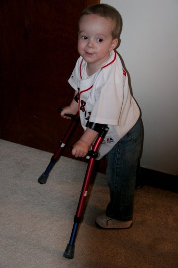
The means of treatment Legg-Perthes-Calvé disease are selected based on the age of the child and the extent of necrosis. In most cases, they consist of immobilizing the head of the femur against the acetabubulum. The leg must be kept in external rotation during these treatments. Several methods allow the specialist to obtain immobilization of the hip.
Prolonged bed rest will force the child to stay still and avoid mobilizing their hip joint.
If the child has trouble staying still, then a splint will be placed.
Plaster is also an often effective means of immobilization.
Fitting a hip orthosis
Femoral or pelvic osteotomy
Physiotherapy: Its objective is to maintain peri-articular muscle volume, and therefore to avoid atrophy.
The use of drugs such as bisphosphonates, could protect the bones against their destruction but their study is still in progress.
Total hip replacement (hip prosthesis) in early adulthood is a solution that can be offered in some cases. This avoids a hip osteoarthritis long-term.
Tracking
La Legg-Perthes-Calvé disease is a pathology whose evolution is unpredictable. It is therefore incumbent on the specialist to carry out regular follow-up in the child carrying this pathology at the beginning stage.
He will have to perform periodic X-rays to detect as early as possible a complication that would require different treatment. This follow-up will take place until bone maturation and definitive surgical treatment will be proposed.
The psychological aspect must also intervene in order to explain to the child why he should not do certain sports and other activities like other children his age.
Conclusion
The disease of Legg-Perthes-Calve ou primary osteochondritis of the hip (OPH) is a pathology that affects children between 02 and 12 years old. It is due to necrosis of the femoral head following a defect in the vascularization of the bone tissue. The causes of this disease are not well known, but certain risk factors have been identified.
La illness ofLegg-Perthes-Calve manifests as lameness, groin pain and limitation of hip movement. The standard radiograph is the main means of confirming the diagnosis but also shows signs of advanced evolution possibly.
Treatment is based more on the principle of immobilization of the hip joint by various orthopedic and surgical means. Management must be early and adequate in order to avoid coxarthrosis (arthrosis of the hip). The psychological care of the child is also very important.

")

")

")

")
