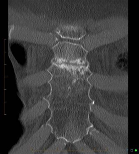
If you feel a chest pain following a violent impact on your chest, it would be best to consult a medical emergency as soon as possible.
Although rare, the sternum fractures can accompany a chest trauma following a car accident or a violent shock to the rib cage.
This article will discuss the semiology of sternal fractures, their causes, as well as the different therapeutic options to treat it.
Sternum fracture: what is it?
There’s nothing quite like a fracture is defined by the presence of a solution of continuity on a os, most often following a direct or indirect trauma (fall, accident, twist, etc.).
There’s nothing quite like a sternum fracture is a lesion of the bone located on the midline of the anterior part of the rib cage : the sternum.
Although it is most often of origin traumatic, it is worth noting that the sternum fracture occurs more easily in subjects over the age of 60, often affected by bone fragility due toosteoporosis (decreased bone mass).
Anatomy of the rib cage
The rib cage (from the Greek thorax, chest) represents the osteo-cartilaginous framework made up of the bones of the thorax which participates in the protection of multiple vital organs: heart, lung, thoracic aorta, etc.
She understands :
- Le sternum which is a flat, odd-numbered bone, located in the center of the chest.
It is made up of three parts (from top to bottom):
- The sternal manubrium
- The body of the sternum.
- The xiphoid process.
- The thoracic (or dorsal) spine
located behind, is composed of 12 vertebrae, themselves separated by the intervertebral discs.
- There’s plenty of ribs
24 in number (12 on each side) are long and curved bones starting from the rachis and articulating at the sternum thanks to the costal cartilage, with the exception of the last two ribs named floating ribs.
The whole forms the chest cavity which contains the two lungs, the mediastinum (space located between the two lungs) which houses the heart, the trachea, esophagus as well as several lymphatic and blood vessels.
What causes a sternal fracture?
The vast majority of sternum fractures are caused by direct trauma to the chest.
In this case, two types of sternum fractures :
- isolated: the rarest, consequences of a direct impact on the sternum and are usually due to car accidents related to the seat belt or the practice of violent sports.
- Secondary to polytrauma: correspond to 2/3 of the sternal tears. They will frequently be associated with fractures of the costal arches and collarbones.
Furthermore, fractures of the sternum can be the result of bone fragility induced by certain pathological states, namely: menopause, vitamin deficiencies et calcium. All of these factors result in a decrease in bone mass, resulting inosteoporosis.
How to recognize a broken sternum?
It will be mentioned before:
- There’s nothing quite like a pain vivid and exquisite, very precisely located at the level of the fracture site.
- There’s nothing quite like a exacerbation of pain during respiratory movements, coughing and sneezing
- Un edema next to the fracture point.
- Un hematoma or an bruise.
Diagnosis and complementary examinations
There’s nothing quite like a sternal fracture will be suspected either in the face of a specific semiology, or in the event of the presence of complications related to the displacement of the fracture or the presence of other associated fractures.
La dyspnea (breathing difficulty) associated with anterior pain spontaneous or triggered by pressure on the sternum with decreased chest expansion are suggestive of a chest fracture.
Added to this the context of trauma (road accident or other.) with the presence of hematomas, bruises and peri-thoracic edema next to the sternum.
The association of a sternal fracture with vertebral or costal lesions is possible. In this context, visceral lesions can be induced: pulmonary contusion, section of the aorta, pneumothorax, pleural effusion, thoracic flap, etc.
What exams to perform?
To confirm the presence of a displaced fracture or not, additional examinations will be requested.
Chest X-ray
In first intention, a standard imagery carried out from the front will make it possible to visualize fractures of the sternum with or without displacement.
It also allows the visualization of pleuropulmonary complications, such as pleural effusions (accumulation of fluid in the pleural cavity) and pneumothorax (abnormal presence of air in the pleural cavity).
Scanner or CT
Is the first-line examination to be carried out in the event of thoracic trauma with or without suspicion of polyfractures.
This examination allows a better visualization of the bone lesions as well as the highlighting of the associated complications, provided that the patient is stable.
What about therapeutic means?
As a general rule, no surgical treatment is required for an isolated and benign fracture of the sternum. Nevertheless, in rare cases (complications) an intervention will be necessary in order to restore bone continuity.
The therapeutic components used are:
Conservative treatment
Faced with a fracture without displacement, that is to say a fracture where the broken bone fragments are still in place, we recommend repos during about 04 weeks, which corresponds to the average time to union of a non-displaced fracture of the sternum.
Rest will be associated with drug treatment aimed at relieving pain and associated inflammatory signs.
The prescribed treatment is mainly based on taking analgesics and anti-inflammatories (NSAIDs).
Surgical treatment
Surgery will be considered if:
- Fractures with displacement.
- Comminuted fractures which are complete fractures in which the bone is broken into multiple fragments.
- Absence of consolidation of the bone.
THEsurgical intervention consists in implanting a material ofosteosynthesis which aims to fix the bones in their original anatomical position in order to allow good consolidation.
It will allow a realignment of the sternum in the axis manubrium – body – xiphoid process.
If you have a complicated fractures it will be necessary to consider:
- Un chest drainage pleural effusions (accumulation of fluid in the pleural space).
- There’s nothing quite like a osteosynthesis
- The treatment of myocardial or pleuropulmonary lesions following a thoracotomy (incision allowing the opening of the chest wall) or a sternotomy (surgical opening of the sternum) completing the fracture.
The delay of consolidation a cas de sternal surgery is on average 06 to 08 weeks.
The daily wearing of a chest vest allows a good consolidation.
In the absence of complications or pathological context, sternal fractures are fractures that consolidate very well, without there being any functional sequelae.
My name is Sidali. I am a general practitioner and Web Editor. As a healthcare professional, my mission is to contribute to the relief of my patients' ailments. Being also passionate about writing, I have the pleasure of sharing my solid medical knowledge with the greatest number of readers, by writing popular articles that are very pleasant to read.


