
Twelve pairs of nerves, called " cranial nerves “, directly connect the brain to the different parts of our body: the head, the neck and the trunk. Let's find out together what there is to know about these nerves.
Cranial Nerves Anatomy
The cranial nerves are the nerves that emerge directly du brainstem (brain). They have the role of receive information from the organs located in the head, with the exception of the vagus nerves which extend into the thoracic and abdominal cavities.
There are 12 pairs, numbered from I to XII, most of which are motor nerves et sensory.
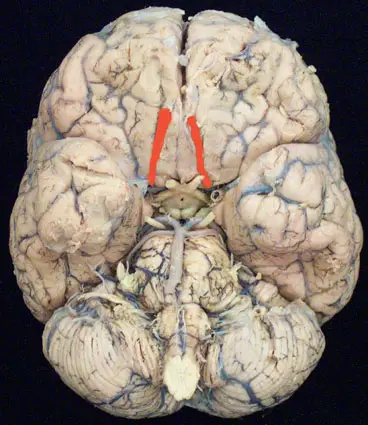
Olfactory nerve (I)
The olfactory nerves emerge from the mucosa of the nasal cavities. The nerve fibers that constitute them come directly from the nasal fossae then cross a horizontal bone plate located above (cribriform plate of the ethmoid) in order to reach the olfactory bulb. This is located just at the base of the skull. All the information that will arrive at this region will then propagate to the temporal lobe, the frontal lobe and the cortex. This last structure plays an important role in memorization.
As we have two nasal cavities, there are then two olfactory bulbs: the left olfactory bulb and the right olfactory bulb.
The olfactory nerves are the cranial nerves The shorter. They carry sensory information me on odor.
Optic nerve (II)
Le optic nerve originates in the papilla (small disc located on the retina). The nerve thus formed crosses the orbital cavity. It enters the interior of the skull through the optic canal, then joins the second optic nerve, originating from the other eye. The 2 optic nerves take an oblique direction towards the back and the interior then merge to form the optic chiasm.
The optic nerve has the sole role of transmitting the images perceived by the eyes to the brain.
Common ocular motor nerve (III)
It’s a motor nerve responsible for eye movement. It starts from the brain stem to join the muscles of the eye as well as the muscles that operate the upper eyelids. A nucleus in the brainstem coordinates these muscles.
This nerve performs various functions:
- muscle innervation here carry the eyeball en in and adduction (allows you to look within);
- control of the levator muscle of the upper eyelid (allows the opening of the eyelids);
- control of the constrictor muscle of the iris, which is also the constrictor of the pupil.
Trochlear nerve (IV)
Ce motor nerve part of the midbrain (upper part of the brainstem). It rises forwards (upward path) to reach the orbital cavity.
Il control le superior oblique eye muscle who ensures the eye rotation to socks and towards theinterior.
Trigeminal nerve (V)
Le trigeminal nerve is the cranial nerve with the greater number of neurofibers and therefore of the greatest transverse diameter. It's a mixed nerve, that is to say both motor and sensory. It starts from the annular pons (middle part of the brainstem).
He's responsible of the skin sensitivity of the face and its cavities. Its name comes from the fact that it is form by meeting of three branches main: the ophthalmic nerve, maxillary nerve, mandibular nerve. The motor nerve controls the chewing muscles and manages the production of saliva and tears. The sensory nerve provides sensitivity to almost all the skin of the face, scalp, teeth, oral cavity, upper eyelid, sinuses, and anterior two-thirds of the tongue.
External ocular motor nerve (VI)
Le external ocular motor nerve originates in the brainstem and terminates in the lateral muscle within the bony orbit. He allows the eyes de look to the side. It activates the levator muscle of the eyelid, as well as the muscles that control the curvature of the lens.
Facial nerve (VII)
Le facial nerve emerges from the brain stem in the sulcus between the pons and the medulla oblongata.
Functionally, the facial nerve is primarily a motor nerve for the muscles of the face and therefore responsible de l 'facial expression. It is accompanied by the intermediate nerve called Wrisberg », sensory nerve for anterior two-thirds taste receptors of the language and also including a small sensitive territory au external acoustic meatus (ear).
Auditory or vestibulo-cochlear nerve (VIII)
The vestibulo-cochlear nerve is a sensory nerve carrying auditory information from the cochlear nerve and information for maintaining balance (upper and lower vestibular nerves).
The vestibulocochlear nerve arises from the brainstem. It is made up of two nerves that run side by side, the cochlear nerve and the vestibular nerve.
The cochlear nerve runs from the cochlea to the brainstem. The information is then transmitted to the auditory center of the temporal lobe of the brain.
While the vestibular nerve runs from the inner ear (vestibule) to the brainstem, nerve connections exist with the cerebellum.
Glossopharyngeal nerve (IX)
The glossopharyngeal nerve is a typically mixed nerve, comprising motor fibers for pharyngeal muscles. It also features sensory neurofibers for posterior part of the tongue and sensory fibers for bucco-pharyngeal region. Finally, it contains parasympathetic fibers intended essentially for the parotid gland.
Vagus nerve or vagus nerve (X)
The vagus nerve, or pneumogastric nerve, is the longer opportunities, cranial nerves. It's the single cranial nerve here innervates opportunities, organs outside the head and neck. In addition, it is the nerve that controls the parasympathetic system in the direction of the heart and the digestive system.
It emerges from the medulla oblongata (part of the brainstem) and innervates, through its motor fibres, the soft palate and the pharynx. As for its vegetative fibers (the parasympathetic), they innervate the heart, the trachea, the lungs, the esophagus, the stomach and the intestines. It is therefore a nerve that participates in the control of swallowing, breathing, circulation and digestion.
Ce nerf at a time sensory and motor is able to release acetylcholine, which causes the bronchi to contract or the heart to slow down.
Spinal nerve (XI)
It’s a motor nerve particular, because it is both cranial and spinal, part of its fibers come from the brain stem (brain) and another part from the spinal cord (spinal root).
Il innervates, for its cranial part, the soft palate muscles and larynx (laryngeal nerve) and for its spinal part, the sternocleidomastoid muscle (on the side of the neck) and the trapezius muscle (behind the neck and shoulder), which participate in the movements of the head and neck.
Greater hypoglossal nerve (XII)
It's the motor nerve of the language. It starts from the medulla oblongata (medulla oblongata) and travels to the base of the tongue, whose movements it controls. He plays a role in the swallowing, but also in the speaking.
Cranial nerve disorders
Olfactory nerve (I)
Damage to this nerve can cause partial abolition (hyposmia) or complete de l 'smell (anosmia).
Optic nerve (II)
Le optic nerve may be the seat ofinflammatory conditions (optic neurites), vascular, toxiques, tumorous ou degenerative.
Common ocular motor nerve (III)
Damage to this nerve can lead to ptosis (drooping upper eyelid), a diplopia (double vision), an abnormal deviation of the visual axis of one eye relative to the other (strabismus), a mydriasis (dilation of the pupil), and accommodation disorders (discomfort with near vision).
Trochlear nerve (IV)
The first sign of trochlear nerve damage is diplopia (we see double), the globes do not move exactly synergistically.
Trigeminal nerve (V)
Trigeminal neuralgia causes pain very intense and facial tics.
External ocular motor nerve (VI)
It is often harmed during the head trauma (fracture of the base of the skull) or compressed by an intracranial tumor. The most common manifestations of its impairment are diplopia (double vision) or a strabismus (abnormal deviation of the visual axis of one eye relative to the other).
Facial nerve (VII)
A facial nerve injury (due to an infection, or a cerebrovascular accident, etc.) causes a facial paralysis : on the affected side of the face, the skin is flabby, without wrinkles, without folds, the eyelids do not close completely, you can no longer smile; on the intact side, the commissure of the lips is retracted. Sometimes damage to the facial nerve can lead to loss of taste sensation (taste).
Auditory or vestibulo-cochlear nerve (VIII)
Damage to the cochleovestibular nerve leads to hearing loss (deafness) and a balance disorder. The most common causes are ototoxic drugs (aminoglycoside antibiotics), or compression by a tumor such as a neuroma. Meningeal infections (meningitis, encephalitis) when not controlled are also responsible for hearing damage.
Glossopharyngeal nerve (IX)
Neuralgia of the IX is most often idiopathic (without known cause) or linked to a vascular-nervous conflict. It can rarely be due to shingles. The pain is of type dazzling, sitting on the base of the tongue and lateral wall of the pharynx. It can be accompanied by a vagal hypertension paroxysmal with syncope vasoplegic.
Pneumogastric nerve (X)
Overactivity of the vagus nerve can trigger a loss of consciousness (vagal syncope), a increase opportunities, gastric acid secretions (causing a peptic ulcer), etc. …
Spinal nerve (XI)
Spinal nerve damage is rare. They are mostly original. traumatic. Damage to this nerve can lead to sternocleidomastoid palsy or trapeze.
Greater hypoglossal nerve (XII)
Disorders of the hypoglossal nerve cause tongue paralysis, usually on one side.
References
https://www.neuromedia.ca/quels-sont-les-12-nerfs-craniens/
https://p1.asso2atp.fr/wp-content/uploads/2019/11/CH8Les-nerfs-cra%CC%82niens.pdf
https://www.edimark.fr/Front/frontpost/getfiles/1832.pdf
https://anatomy.elpaso.ttuhsc.edu/modules/CN_module/cnI.html

My name is Arotoky and I am studying human medicine. Web editor for more than 5 years, I currently focus on the theme of health and well-being. I have the strong conviction that with my valuable articles, I can help many people to relieve their ailments and feel better. My goal is to share my health knowledge with the general public through web writing.