
A very rare phenomenon, craniosynostosis designates a cranial deformation which concerns approximately one birth in 2500. It is generally observed from the birth of the baby or in the first months following childbirth. In addition to the aesthetic consequence, this disease also has serious consequences on the health and growth of the child. It is therefore important to take care of it without delay.
What is craniosynostosis ? What are its causes? What complications can occur? How to treat this disease? We will take stock in this article.
Definition and clinical presentation
Craniostenosis is a cranial deformity that affects newborns. It affects the skull, face and sometimes other organs (brain, eyes, nose and ears).
At birth, some areas of the baby's skull are soft. They are called fontanelles. It is at these fontanelles that the bones of the skull join, but do not yet fuse together. They are held together by cartilage called sutures. They are found at the level of the frontal bone, between the forehead and the rest of the skull, between the parietal bones and at the back of the skull. The presence of these sutures facilitates the passage of the baby's head at the time of delivery and the development of the brain afterwards.
La craniosynostosis manifests itself differently depending on the suture area concerned.
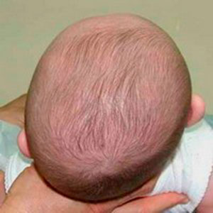
In the majority of cases, there is ossification of the sagittal sutures, between the two parietal bones. That's what we call scapocephaly. This leads to an increase in the length of the skull, because it cannot grow in width. Therefore, the baby may have either a bulging forehead or a bump on the back of the skull. The scapocephaly is more common than other types of craniosynostosis (in 50% of cases). It affects more boys than girls.
When the craniosynostosis concerns the coronal suture, that is to say on the transverse joint between the frontal bone and the parietal bones, we speak of the plagiocéphalie. It is characterized by an asymmetrical shape of the baby's skull. It gives the head a "flat" shape.
To know everything about plagiocephaly, see the following article.
Craniostenosis can also sometimes affect the metopic suture, the one located at the level of the forehead. In this case, we are talking about trigonocephaly. The result is manifested by the development of a kind of bony ridge, a triangular forehead and close-set eyes.
In general, the diagnosis of craniosynostosis is clinical. It can be placed during the first days of the baby's life through inspection and palpation of the skull. So the doctor suspect craniosynostosis in the presence of a linear bead at the level of the junction of two bones of the cranial box. However, in some cases, the diagnosis is late. More rarely, this malformation is detected a little early in pregnancy, during a scan.
In order to confirm craniosynostosis and develop a strategy for the treatment of the disease, it is often useful to resort to radiological examinations. Regardless of whether the malformation is non-syndromic or syndromic, a spiral CT scan with a 3D (three-dimensional) reconstruction makes it possible to clearly visualize the characteristics of craniosynostosis. The scanner is a reference examination for this type of disease.
What can cause craniosynostosis?
There are two types of craniosynostosis which are syndromic craniosynostosis et non-syndromic craniosynostosis.
La syndromic craniostenosis is caused by genetic abnormalities such as Crouzon's, Apert's or Pfeiffer's syndromes. This type of craniosynostosis occurs rarely (20% of cases).
La non syndromic craniosynostosis, as for it, is not related to any disease. It is said to be "isolated". It is usually related to the early closure of the baby's skull bones during pregnancy or shortly after birth. The real reasons for this phenomenon, however, remain unidentified until now.
La non-syndromic craniosynostosis is the most frequently observed form.
Possible complications of craniosynostosis
Craniostenosis should never be taken lightly. If the disease is delayed in treatment, it can be very dangerous for the baby. Beyond aesthetic discomfort, the major complication of craniosynostosis is intracranial hypertension (HTIC). It results in an increase in intracranial pressure. Indeed, when the skull closes when the brain develops, it is quite normal for it to be compressed. The pressure of the cerebrospinal fluid in the cranium becomes more important.
A intracranial hypertension can lead to headaches, vomiting, cognitive impairment, eye problems and dizziness in the baby.
Le risk of intracranial hypertension increases with the age of the child and the number of sutures involved. Most often, it appears between the ages of 3 and 8 years (in 10% of non-op childrenered).
What treatment to cure craniosynostosis?
Since it is the closing a cranial suture too early which gives rise to the deformation of the skull, the logical solution consists in the reopening of this pathological suture. This is done by a surgery. It is also the only existing treatment for craniosynostosis.
Each form of craniosynostosis uses different techniques. However, the objective remains the same: to reshape the baby's skull in order to recreate a better morphology and allow the proper development of the underlying brain.
The operation consists of cutting, removing and then replacing certain cranial bones. To hold the assembly together, doctors use wires or plastic plates held together by small rivets. For patients less than one year old, absorbable materials can be used.
These are very delicate acts and everything must be done with precision.
This procedure is performed under general anesthesia by a medical team made up of cranio-maxillofacial surgeons and pediatric neurosurgeons. Of course, a consultation of the baby's medical check-up must be done beforehand. A scanner can be done to confirm the closure of the incriminated suture, to ensure the absence of another welded suture and of an intracranial anomaly. It also makes it possible to take measures in order to be able to correct the malformation.
To reduce the risk of complications, the ideal is to perform the operation between 3 and 9 months after the birth of the baby. It is also necessary that the baby weighs at least 6 kilos to be able to be operated. It is from this weight that a child has sufficient blood mass to allow him to undergo an operation in complete safety.
The duration of hospitalization often lasts between 7 and 10 days. After the intervention, the follow-up must be regular: every year then every 2 years, until the end of the growth of the child. It is a medico-surgical, psychological and school follow-up.
Very impressive, this operation causes no sequelae in the child. There may be a scar that goes from ear to ear, but it will soon be hidden in her hair. It also turns out that a child operated on for craniosynostosis is quite fit to perform any sports activity, without contraindication.
To conclude, craniosynostosis is a rare condition that can greatly affect a baby's growth. Cranial surgery is the best, if not the only option to treat it. However, to obtain better morphological results, diagnosis and management must be done as early as possible.
References

My name is Arotoky and I am studying human medicine. Web editor for more than 5 years, I currently focus on the theme of health and well-being. I have the strong conviction that with my valuable articles, I can help many people to relieve their ailments and feel better. My goal is to share my health knowledge with the general public through web writing.