Article reviewed and approved by Dr. Ibtissama Boukas, physician specializing in family medicine
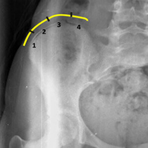
The Risser test is used in medicine to assess the skeletal bone maturity based on the level of ossification and fusion of the iliac crest processes. In particular, it makes it possible to guide treatment in children with scoliosis, but also other pathologies related to the growth plate (such as Osgood-Schlatter disease).
To know everything about the scoliosis in children (including diagnosis and management), see the following article.
Basically, if growth is complete, treatments such as scoliosis corsets are less likely to prevent spinal deviations. It will then be necessary to consider a surgery if the deviation is significant and incapacitating. On the contrary, if the child is in full growth period, it will be necessary to correct the deviation in an aggressive way (for example using a adapted corset), and thus prevent the progression of the disease.
Classification
The Risser test is assessed via an AP X-ray of the pelvis to observe the iliac crest. Ideally, this test should be performed under strict conditions and interpreted by radiologists to avoid errors. Here is the detailed explanation and interpretation of the American Risser test classification:
- Stage 0: no ossification at the level of the apophysis of the iliac crest (absence of cartilage)
- Stage 1: cartilage covering less than 25% of the iliac crest; it corresponds to prepuberty
- Stage 2: cartilage covering 25-50% of the iliac crest; it corresponds to the stage preceding or in full growth spurt.
- Stage 3: cartilage covering 50-75% of the iliac crest; it corresponds to the slowing down of growth.
- Stage 4: cartilage covering >75% of the iliac crest; it corresponds to a virtual cessation of growth.
- Stage 5: complete ossification and fusion of the iliac crest apophysis (cartilage completely attached to the iliac crest); it corresponds to the end of growth
As stage 0 and stage 5 may appear similar (given no evidence of ossification), the patient's age and long bone growth plates should be considered to help distinguish between them. A stage 0 patient will still have growth plates in most of their long bones, and will likely be under 16 (for a girl) or 18 (for a boy). A stage 5 patient, on the other hand, will not have growth plates in their long bones and will be older.
Note: In the French system, the classification used to quantify the Risser test is slightly different. More specifically, it has 4 stages (instead of 5) characterized as follows:
- Stage 0: no ossification at the iliac crest apophysis
- Stage 1: ossification and fusion between 0-33%
- Stage 2: ossification and fusion between 33-66%
- Stage 3: ossification and fusion between >66%
- Stage 4: Complete ossification and fusion of the iliac crest apophysis.
alternatives
Although the Risser test is a reliable radiographic parameter to assess the growth potential of children with scoliosis and to determine the skeletal bone maturity, there are other tests that have the same role. Among the most popular are:
- Tanner-Whitehouse-III method
- Atlas of Greulich and Pyle
- Oxford method
- Morphological assessment of olecranon development
To maximize the predictive value of the Risser test, it should be used in conjunction with other tools such as skeletal age, actual patient age, and time since first menstruation in women.
References
- https://radiopaedia.org/articles/risser-classification
- https://www.ncbi.nlm.nih.gov/pmc/articles/PMC3392381/
- https://en.wikipedia.org/wiki/Risser_sign
My name is Anas Boukas and I am a physiotherapist. My mission ? Helping people who are suffering before their pain worsens and becomes chronic. I am also of the opinion that an educated patient greatly increases their chances of recovery. This is why I created Healthforall Group, a network of medical sites, in association with several health professionals.
My journey:
Bachelor's and Master's degrees at the University of Montreal , Physiotherapist for CBI Health,
Physiotherapist for The International Physiotherapy Center


