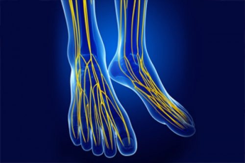
Did you know that the nerf sciatica runs down the leg to the foot? In this article, we are going to look at the simplified anatomy of the foot and discuss the location of the sciatic nerve. Keep reading to find out more!
Lumbar Spine Anatomy
La lumbar spine is a vital part of the human anatomy because it provides structural support to the entire spine. This bony area of the body is located at the base of the spine, just above the sacrum.
The lumbar spine is made up of five vertebrae, each of which is separated by a disc. These discs are responsible for absorbing shock and allowing the spine to move freely.
Between each vertebra emerge nerves which will descend along the lower limbs to provide force and sensation. The sciatic nerve is one of these nerves.
Sciatic nerve : Definition
Le sciatic nerve is the longest and widest nerve in the human body. It starts from the lower back, crosses the buttocks and goes down the back of each leg. the sciatic nerve consists of nerve roots that come out of the lumbar and sacral part of the spine (L4, L5 and S1). It goes to the feet via offshoots. In other words, it branches out into smaller nerves that run down to the toes.
These nerve roots come together to form one large nerve that innervates the legs and feet. the sciatic nerve provides sensation to the skin of the leg and foot, as well as muscle control for movement of the leg and foot.
La sciatica is a condition that occurs when the sciatic nerve is compressed or irritated, causing pain that radiates along the nerve. The treatment of sciatica usually includes exercise, stretching, and physical therapy.
In severe cases, surgery may be needed to relieve pressure on the sciatic nerve.
foot anatomy
Feet are amazing anatomical structures made up of 26 bones, 33 joints, 112 ligaments and tendons, and numerous blood vessels and nerves. These bones are grouped into three categories: tarsals, metatarsals and phalanges.
The tarsals are a group of seven bones that form the ankle joint. The metatarsals are a group of five long bones that run from the ankle to the toes. The phalanges are the bones that make up the joints of the toes. In addition to these bones, the foot also contains a number of muscles and ligaments that help support and move the foot.
The bones of the foot are held together by a network of muscles and ligaments. The muscles of the foot can be divided into two groups: those that act on the ankle joint and those that act on the toe joints. Muscles that act on the ankle joint include the gastrocnemius, soleus, and tibialis anterior.
These muscles work together to plantarflex (orient) the foot. The muscles that act on the joints of the toes are the extensor digitata muscle and the extensor digitata muscle. These muscles work together to dorsiflex (lift) the foot.
Branches of sciatic nerve at foot level
Le sciatic nerve branches into the foot to provide sensation and muscle control for leg and foot movement. The branches of sciatic nerve include:
- Le tibial nerve : The branch of the tibial nerve runs down the calf and into the foot, where it innervates the calf muscles.
- The peroneal nerve: The branch of the peroneal nerve innervates the muscles of the thigh. In the foot, the sciatic nerve branches into several smaller nerves, which innervate the toes.
The pain of sciatic nerve can occur when these small nerves are irritated or compressed. The treatment of pain sciatic nerve usually includes rest, ice, and physical therapy. Sometimes surgery is needed to relieve the pressure on the sciatic nerve.


