
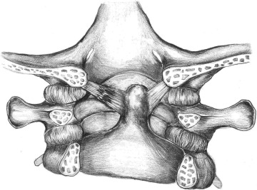
Definition and anatomy
The alar ligaments are ligaments that connect the tooth of the axis (bony projection at the level of the second cervical vertebra C2) to the tubercles of the medial aspect of the occipital condyle. They extend almost horizontally, creating an angle of at least 140° between them.
Located on either side of the skull, these ligaments are short, strong fibrous cords that have the function of controlling the lateral movements of the head when it turns left and right, thus preventing excessive rotational movement. For example, the right alar ligament, when in a stretched position (in cervical flexion), prevents excessive cervical rotation to the left.
Likewise, the anatomical arrangement of the alar ligaments makes it possible to fix the skull to the axis, and stabilize the C1 and C2 vertebrae (especially during cervical rotation).
Clinical assessment
The alar ligaments are susceptible to tearing if force is applied when the head rotates excessively while positioned in flexion.
If an alar ligament is ruptured, the amplitude of rotation of the head relative to the neck increases (over the normal limit of 20 degrees) on the contralateral side, which can lead to cervical instability.
Stability test
Certain clinical tests can be used to assess the integrity of the alar ligaments (and therefore the stability of the upper cervical vertebrae):
Lateral bending test
When performing this test, the therapist stabilizes the spinous process and the spinal blade of the axis (C2 vertebra). Light compression is applied across the skull to facilitate lateral flexion of the atlanto-occipital. Passive lateral flexion is then applied by the therapist, which is equivalent to directing the ear on the side being assessed towards the opposite shoulder.
If fixation of the axis is done properly, no lateral flexion should occur (negative test). It is also recommended to perform the test in 3 planes (neutral, in cervical flexion and in cervical extension) to take into account the variation in the orientation of the alar ligament. A positive test is noted if there is excessive lateral flexion movement in the 3 planes evaluated. This would indicate a probable lesion of the alar ligament.
Rotation test
To carry out this test, the axis (two cervical vertebra, or C2) is fixed by the therapist using a lumbrical grip by stabilizing the laminae and the spinous process of C2. The skull is then gripped and rotated, causing the occiput and atlas (first cervical vertebra, or C1) to rotate with it. As for the lateral flexion test, the test is repeated in 3 positions (neutral, flexion, extension).
Normally, an acceptable rotational amplitude varies between 20 and 40 degrees (test negative), although these limits are subject to certain variations. Laxity in the 3 positions evaluated must be observed to qualify the test as positive. This laxity would suggest a tear of the alar ligament which should ideally be confirmed by a medical imaging examination.
pathology
In addition to traumatic injuries to the alar ligaments (for example during a car accident), these ligaments can be prone to calcifications. However, this condition is relatively rare.
Calcification of the alar ligaments can sometimes cause neck pain and neck pain. This would be explained by calcium deposits at the level of these ligaments. Although magnetic resonance imaging (MRI) and CT scans are better able to identify these calcium deposits, x-rays of the cervical spine also allow their diagnosis.
The differential diagnosis of alar ligament involvement includes the following conditions:
In terms of treatment, it does not differ from conventional cervical damage, and includes the following modalities: relative rest, medication, immobilization if necessary, physiotherapy, infiltration in refractory cases, etc.
References
- https://en.wikipedia.org/wiki/Alar_ligament
- https://www.physio-pedia.com/Alar_Ligament_Test
- https://radiopaedia.org/articles/alar-ligament-calcification?lang=us

My name is Anas Boukas and I am a physiotherapist. My mission ? Helping people who are suffering before their pain worsens and becomes chronic. I am also of the opinion that an educated patient greatly increases their chances of recovery. This is why I created Healthforall Group, a network of medical sites, in association with several health professionals.
My journey:
Bachelor's and Master's degrees at the University of Montreal , Physiotherapist for CBI Health,
Physiotherapist for The International Physiotherapy Center