Article reviewed and approved by Dr. Ibtissama Boukas, physician specializing in family medicine
Leri's sign is used by health professionals when they suspect upper lumbar nerve root involvement. This may come from a cruralgia or an herniated disc, and usually manifests as lower back pain radiating to the groin or the front of the thigh.
Here we explain the Léri test with an emphasis on practical anatomy, and explaining the procedure for this maneuver in a clinical context.
Definition
Léri's sign is used to detect nerve root irritation via passive stretching of the anterior part of the thigh. Contrary to Lasegue test ( SLR) which evaluates the presence of radiculopathy lower lumbar spine, the Léri maneuver assesses the upper lumbar nerve segments. This is also the reason why it is not used as much as the SLR (because the radiculopathies lower back are much more common).
More specifically, the Léri test stresses the crural nerve and upper to middle nerve roots (between L2 and L4). To better understand the technique, let's briefly review the anatomy of the crural nerve.
Crural Nerve Anatomy
Where exactly is the crural nerve located, and what is its course?
Also called the femoral nerve, the crural nerve is composed of nerve fibers coming from the 2nd, 3rd, and 4th lumbar vertebrae (L2, L3, L4). This amalgam of spinal nerves originating from the lumbar plexus passes through the belly, and continues its journey in the leg before dividing into several branches.
The crural nerve is sensory and motor at the same time (also called sensory-motor). In other words, its motor function allows the contraction of certain muscles at hip and knee level (such as hip flexors or knee extensors).
Its sensory function, it allows sensitivity at the level of the front and internal face of the leg and the foot (mainly thanks to the saphenous nerve, one of the most important branches of the crural nerve).
To learn more about the crural nerve and associated pathologies, see the following article.
Procedure
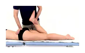
The patient lies prone (on the stomach), and the therapist stands on the affected side. By stabilizing the pelvis to avoid compensations (anterior tilting of the pelvis), the practitioner gradually bends the patient's leg (hip flexion) until the end of the amplitude.
If the test does not elicit any response, nerve tensioning can be continued by raising the leg off the ground (hip extension) while maintaining knee flexion. We can also induce movements of the ankle (plantar flexion) and of the head to put even more tension on the dura mater and the nerve roots.
Léri's sign can also be reproduced in lateral decubitus (when the patient is lying on the side) when the prone position cannot be tolerated. This alternative consists of lying the patient on the unaffected side, then fixing the pelvis while bringing the knee into flexion and the hip into full extension.
Normally, the patient's heel should touch his buttock, and a stretch in the quadriceps should be felt. In the presence of a positive Léri sign, unilateral pain could be reproduced in the lumbar region, buttocks, oldest boy, Or the thigh. In some cases, the pain can even affect the calf, ankle or foot. These symptoms typically appear between 80 and 100 degrees of knee flexion. To improve the specificity of the test, the results should be compared with the healthy side.
A positive Leri's sign may be indicative of a cruralgia from a herniated disc affecting the L2, L3 and/or L4 nerve roots. Typically, pain in the groin and hip region radiating down the medial aspect of the thigh suggests an L3 origin, while pain in the front of the leg indicates a root problem. L4. However, this varies from patient to patient.
Obviously, Leri's sign will be part of a complete examination including a neurological examination and other clinical tests aimed at clarifying the diagnosis. For example, in the case of a herniated disc at the L3/L4 level, there will also be quadriceps muscle weakness associated with an absent or weakened patellar tendon reflex.
To know everything about herniated disc and its management, see the following article.
If the pain in the front of the thigh appears before 80 degrees of flexion of the knee, a contracture or pathology of the quadriceps muscle (rectus femoris) or of the psoas could come into play. Another differential diagnosis indicative of a "false positive" could be a hip disorder.
References
- https://www.physio-pedia.com/Femoral_Nerve_Tension_Test
- https://www.orthofixar.com/special-test/prone-knee-bending-test/
My name is Anas Boukas and I am a physiotherapist. My mission ? Helping people who are suffering before their pain worsens and becomes chronic. I am also of the opinion that an educated patient greatly increases their chances of recovery. This is why I created Healthforall Group, a network of medical sites, in association with several health professionals.
My journey:
Bachelor's and Master's degrees at the University of Montreal , Physiotherapist for CBI Health,
Physiotherapist for The International Physiotherapy Center


