Article reviewed and approved by Dr. Ibtissama Boukas, physician specializing in family medicine
What is the vertebral lamina? This article explains everything you need to know about this component of the vertebral column.
Definition and anatomy of the vertebral body
Before talking about the vertebral laminae, it is worth briefly explaining the anatomy of the spine, and the vertebrae that compose it.
The vertebral column is made up of the juxtaposition of bones called vertebrae. Also called rachis, it is separated as follows:
- 7 cervical vertebrae
- 12 thoracic (or dorsal) vertebrae
- 5 lumbar vertebrae
- 5 sacral vertebrae (forming the sacrum)
- 4 coccygeal vertebrae (fused)
Here is a visual diagram of the spine:
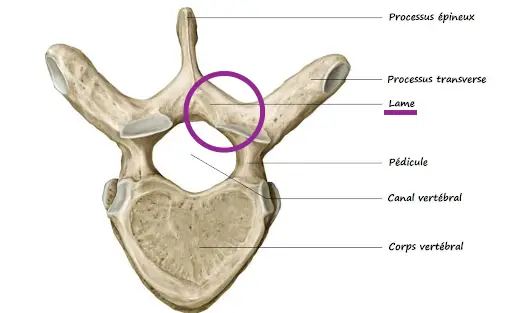
Generally speaking, each vertebra is composed of a vertebral body in its front part, and a posterior arch at the back formed by the pedicles and the vertebral laminae.
The lamina is therefore the part of the vertebra which connects the spinous process (often called spinous process) and the transverse process (or transverse process). There are two laminae per vertebra, located on either side of the spinous process. They are present at the level of cervical spine, dorsal and lumbar.
Pathologies related to the vertebral lamina
The vertebral lamina may be the fracture site.
Treatment related to the vertebral lamina
The spinal lamina is often the site of surgery to relieve symptoms caused by pressure on the nerve roots. This can happen in the case of a narrow lumbar canal, or a herniated disc.
This intervention is called laminectomies


