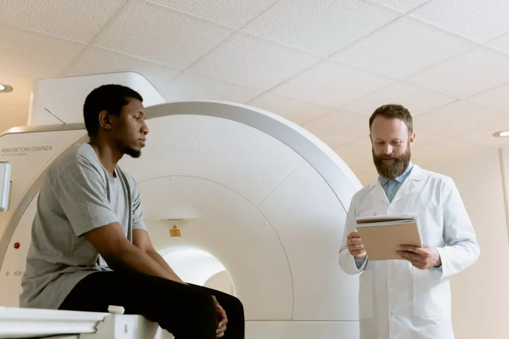
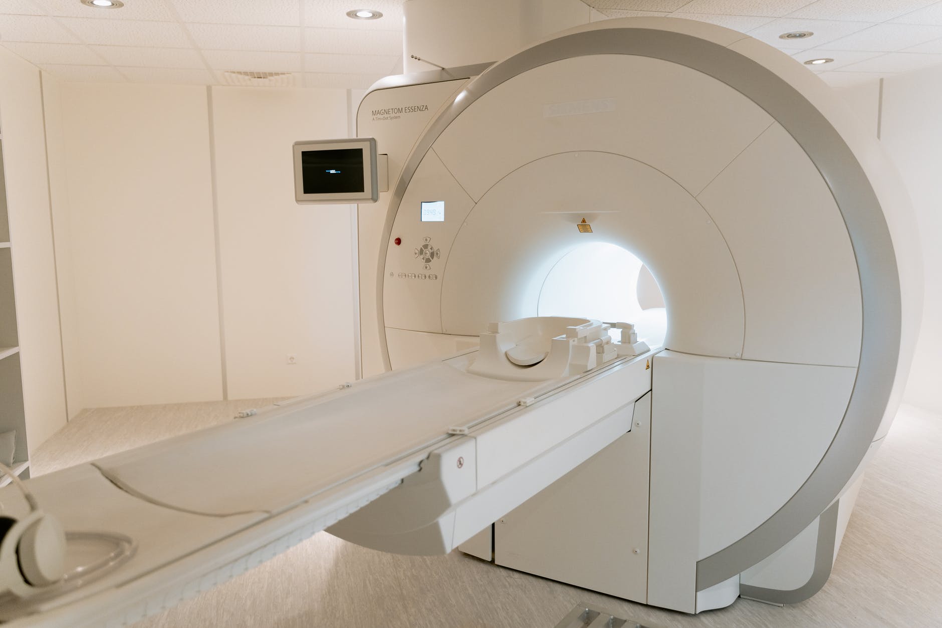
Magnetic resonance imaging (MRI) is a non-invasive medical examination that provides very detailed information on the morphology of the internal structures of the body.
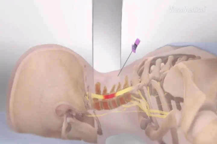
For example, cervical MRI makes it possible to study with precision the various anatomical elements of the cervical region, in particular the spinal cord and its nerve roots, intervertebral discs, the thyroid gland, the upper airways (larynx, trachea)…
For even more precision, your doctor may prescribe a « Cervical MRI with injection ».
So what is the latter? When is it needed? What more does it bring compared to a classic cervical MRI (without injection)? These are the questions we will answer in this article.
Cervical region: anatomical reminder
Le of, or "cervical region", is the part of the body that connects the head to the trunk. It is supported by the cervical segment of the spine, a mobile bony framework around which numerous muscles are superimposed.
The cervical region is limited above by the mandibular arch (lower jaw) and below by a plane passing through the upper part of the sternum (sternal manubrium).
The main bony element of the neck is obviously the cervical spine. This mobile segment of the spine , is made up of 7 vertebrae cervical numbered from C1 to C7.
Each vertebrate is made up of a vertebral body, of two transverse processes laterally and from posterior vertebral arch giving a spinous process visible and palpable on the back of the neck.
Each vertebra is dug in its center with a hole called « vertebral foramen ». The stacking of these vertebral foramina forms the spinal canal containing the spinal cord and its various elements.
For better flexibility, mobility and shock absorption, there is a kind of fibrocartilaginous cushion between the vertebrae cervical called intervertebral disc.
In addition to the spine (and its nervous content), the cervical region is very rich in primordial anatomical elements:
- Muscular elements: the cervical region contains many muscles. We can mention in particular the muscle hider of the neck, the sternocleidomastoid (SCM), the trapeze, splenius head and neck back, semi-thorny head and neck, multifid, scalenes, muscles above et subhyoid...
- Tendon elements: one can, among other things, see projecting at the level of the sternal manubrium (upper part of the sternum) the two tendons of the SCM (sterno-cleido-mastoid) muscles forming what is called the sternal notch.
- Organic elements: one of the most important glands in the human body, the thyroid gland, as well as parathyroid. We also find the upper parts of the digestive tract and the respiratory system such as the esophagus, trachea and larynx.
- Vascular elements: at the level of the cervical region run large-caliber vessels intended in particular to irrigate the brain. We can cite the subclavian arteries, internal carotids et external, vertebral arteries, internal and external jugular veins.
Pathologies that may affect the cervical spine
Le cervical spine can be the seat of a wide variety of pathologies:
- The vertebral fractures : they can be post-traumatic or without notion of trauma (pathological fractures).
- The cervical sprains : injury to one or more ligaments that hold the cervical vertebrae together.
- THEcervical spondylosis ou cervicarthrosis: wear of the cartilage at the level of the intervertebral joints of the cervical spine.
- Cervical arthritis: inflammation of the joints of the cervical spine, particularly in the context of rheumatoid arthritis.
- Abnormalities of cervical lordosis: accentuation of cervical lordosis or “cervical hyperlordosis”; attenuation of physiological cervical lordosis or « hypolordosis cervical » ; straight aspect of the cervical spine or « straightness of the cervical spine », reversal of cervical lordosis or « cervical kyphosis ».
- La cervical scoliosis : deformation in the frontal plane of the cervico-dorsal spine (rarer than thoraco-lumbar scoliosis, but it does exist).
- La cervical spinal stenosis : narrowing of the cervical canal.
- THEspinal cord infarction : spinal cord vascular accident (the spinal cord is no longer properly vascularized).
- The abnormalities of intervertebral disc neck: degeneration and herniated disc cervical, inflammatory disc disease…
To make one of these diagnoses, the doctor first performs a physical examination comprising a careful questioning and exam complete physical in order to collect as much information as possible about the patient (history, symptoms and their characteristics, circumstances of a possible accident, highlighting of certain physical signs, etc.).
Often (but not always), the doctor will order a exam complementary to support his diagnosis. He may, for example, resort to an imaging examination such as plain x-ray cervical spine, computed tomography (scanner) ou theIRM.
What is cervical MRI?
A IRM, or " Magnetic resonance imaging ", is one of the techniques ofmedical imaging latest and perfected. It allows you to visualize the different anatomical elements of the body with great precision and in a non-invasive way.
Unlike standard radiography or computed tomography (scanner), MRI does not use x-rays but a magnetic field. The latter is perfectly harmless, which makes MRI feasible even in pregnant women.
THECervical MRI is an MRI that explores the neck region for diagnostic purposes. Above all, it allows you to explore the soft tissue such as spinal cord, the nerve roots of the latter, the intervertebral discs and the thyroid gland, unlike the scanner which better studies the bone structures.
Contrast agent associated with MRI: why?
Due to the heterogeneity of the different tissues of the body, MRI allows the visualization of fairly detailed images by obtaining a spontaneous contrast. It is therefore possible to study organs, cardiac chambers and vessels with sufficient precision without necessarily having to resort to the injection of a contrast product.
To obtain more precise and better quality images, it is often necessary to increase the contrast of the different tissues by intravenous injection of a contrast agent (PDC).
What is a contrast product?
Un contrast agent (PDC) is therefore a molecule intended to improve the quality of MRI diagnostics by obtaining more detailed shots.
Generally, products based on gadolinium (chemical element whose atomic number is 64), because the latter is spontaneously visible on MRI. In rare cases (allergy to gadolinium for example), it is replaced by contrast products based on diode.
These contrast agents are injected by venous route after placing a catheter in a vein (usually a vein in the arm). They then pass through the general bloodstream before reaching the area to be explored to opacify it.
When is it necessary to use PDC?
It is up to your doctor to decide on the type of MRI to perform depending on the organs to be explored or the suspected pathology. For example, exploring the vascular structures of the neck or looking for a cervical tumor will almost systematically require an MRI with injection of PDC.
It happens that the radiologist in charge of carrying out the MRI begins with an examination without injection before deciding, according to the images that he visualizes on the control screens (in real time), of the relevance of an injection of PDC.
In other words, if a simple MRI was sufficient to highlight the diagnosis sought, it will not be necessary to inject PDC!
Cervical MRI with injection of PDC: what does it bring more compared to an MRI without injection?
MRI with injection of PDC makes it possible to obtain isharper and more detailed images cervical anatomical structures, especially those made up of soft tissue. It therefore proves to be a valuable aid in the diagnosis of many pathologies affecting the cervical region.
However, injection of a contrast medium in the body is not a totally innocuous gesture. It involves many risks and has many contraindications. This is why, before prescribing this type of examination, your doctor will carefully assess his benefit/risk ratio and will ensure that there are no contraindications (kidney failure, allergy, pregnancy, etc.).
Projects
[1] “Nuclear Magnetic Resonance Imaging (MRI) – Special Topics”, MSD Manual Professional Edition. https://www.msdmanuals.com/en/professional/special-topics/principles-of-radiological-imaging/magnetic-resonance-imaging-nuclear-mri (accessed August 3, 2022).
[2] S. Lee, “Magnetic Resonance Imaging (MRI)”, Canadian Cancer Society. https://cancer.ca/en/treatments/tests-and-procedures/magnetic-resonance-imaging-mri (accessed August 4, 2022).
[3] “How is an MRI performed? » https://www.ameli.fr/assure/sante/examen/enseignement-medicale/deroulement-irm (consulted on August 4, 2022).
[4] "MRI (nuclear magnetic resonance imaging)", VIDAL. https://www.vidal.fr/sante/examens-tests-analyses-medicales/irm-images-resonance-magnetique.html (accessed August 4, 2022).
My name is Katia, I am specialized web editor in writing medical articles. Being passionate about medicine and writing, I set myself the goal of making medical information accessible to as many people as possible, through the popularization of even more complex scientific concepts.

