Article reviewed and approved by Dr. Ibtissama Boukas, physician specializing in family medicine
A laminoplasty is a surgical procedure to widen the spinal canal. It is mainly carried out at the level of cervical spine. It can sometimes be performed at the thoracic or lumbar level, especially in children or young adults.
This article tells you everything you need to know about laminoplasty.
Definition and anatomy
To better understand laminoplasty, let's discuss the anatomy of the vertebrate, and more specifically the anatomical location of the spinal blades.
Generally speaking, each vertebra is composed of a vertebral body in its front part, and of a posterior arch at the back formed by the pedicles and the vertebral laminae.
The lamina is therefore the part of the vertebra which connects the spinous process (often called spinous process) and the transverse process (or transverse process). There are two laminae per vertebra, located on either side of the spinous process. They are present at the column level cervical, dorsal et lumbar.
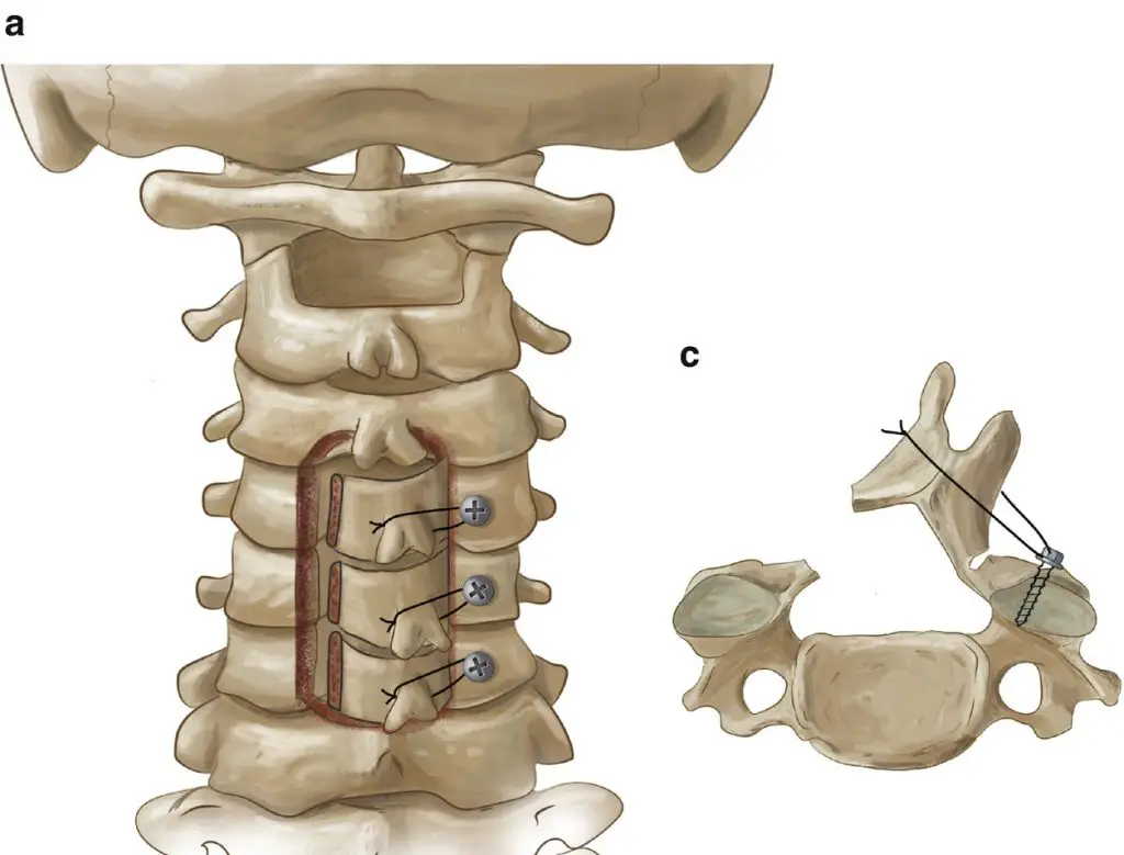
In laminoplasty, one or more portions of the blade are removed to widen the spinal canal. The blades are then reshaped or repositioned to decompress the spinal cord and spinal nerves. Metal rods, an implant or a bone graft are also used to maintain the enlarged space.
Difference with laminectomy
A procedure similar to laminoplasty is laminectomy. The latter removes the vertebral lamina outright instead of creating an opening.
Compared to laminectomy, which is a more complex surgery, laminoplasty leaves a larger part of the bone and ligament structure intact. This is essential to maintain normal spinal anatomy and curvatures, as well as to reduce the risk of complications (Source).
Laminoplasty is more appropriate for children and young adults. Indeed, pediatric patients have a higher risk of spinal instability following laminectomy compared to adults.
However, since the laminoplasty preserves the anatomy of the posterior arch, it allows less decompression. Thus, people who require significant decompression because of their condition must go through laminectomy at all costs.
Other types of decompressive surgery
Alternatives to laminoplasty include:
- Laminectomy
- Discectomy
- Corpectomy
- Foraminotomy
- Arthrodesis
- Flavectomy
It should be noted that the optimal surgical technique to treat spinal or nerve root compression remains controversial. One approach is not necessarily superior in all circumstances, and the best option will depend on patient-specific anatomical and symptomatic factors.
Indication
Laminoplasty is usually done to correct conditions where the spinal canal is narrowed, putting pressure on the spinal nerves or spinal cord. It is generally when the symptoms become severe and incapacitating despite conservative treatment that surgery is considered.
Here are the conditions that may benefit from a laminoplasty:
- Myelopathy
- Narrow lumbar canal (video explanation)
- Herniated disc
- Degenerative disc disease
- Tumors
- Traumas and fractures
- Vascular malformations
- Syringomyelia
- Yellow ligament hypertrophy
- Spondylolisthesis
In general, the observed symptoms that alert the medical team are the following:
- Severe pain with irradiations
- Severe and incapacitating paresthesias
- Upper and lower limb weakness
- Gait disorder
- Balance issues
- Incontinence
- Coordination disorder
Procedure
Laminoplasty is a surgical procedure performed under general anesthesia. In the case of a cervical laminoplasty (the most common), an incision of about 7 to 10 cm is made in the middle of the neck, and the back of the spine.
Using an operating microscope and other surgical instruments, the surgeon will essentially free up space in the spinal canal, thereby reducing pressure on the spinal cord and spinal nerves. The opening of the canal will be maintained by a small metal plate or a bone graft.
To make sure not to irritate the spinal cord and spinal nerves, the intervention will often be carried out under control of the evoked potentials. Basically, we want to make sure that there is no damage to the nerve structures during the operation.
To do this, we will place electrodes on the scalp and on the legs. During the surgery, we will regularly send a weak electric current through the electrodes to ensure the integrity of the spinal cord.
At the very end of the operation, the surgeon closes the incision and bandages it with a bandage after cleaning the wound.
Recovery and recovery
Patients usually spend the night in the hospital after the operation, and are discharged the following day or the day after.
In the majority of cases, the surgeon will not recommend wearing a cervical collar after a laminoplasty. On the other hand, some patients prefer to wear a flexible collar to support the cervical spine during the days following the operation. In children, a rigid collar may be prescribed for up to 6 weeks.
Personalized painkillers and/or muscle relaxants will usually be prescribed to control pain and reduce muscle tension. The medication will then be weaned off gradually.
A follow-up visit will usually be fixed 4 to 6 weeks after surgery. X-rays will be taken to observe progress, monitor healing and ensure bone fusion.
Overall, it is estimated that the total recovery period is 6-8 weeks for an corpectomy.
Re-education
Patients are generally encouraged to increase their level of physical activity as tolerated, while refraining from strenuous exercise (eg, activities involving jumping).
To optimize rehabilitation, the doctor may prescribe physiotherapy (physiotherapy). This will speed healing, and promote a safe and effective recovery. The methods used will be:
- Electrotherapy
- Analgesic modalities (heat, ice, etc.)
- Gentle massage
- Soft mobilizations
- Therapeutic exercises
- Postural exercises
- Advice and education
- Etc.
Complications
Although relatively rare, complications following a laminoplasty can include:
- neurological deficits due to nerve damage
- loss of spinal alignment
- spinal cord damage
- damage to the trachea or esophagus
- wound infection
- bleeding
- displacement of the graft
- vertebral artery injury
Source
- https://www.ncbi.nlm.nih.gov/pmc/articles/PMC7154346/
My name is Anas Boukas and I am a physiotherapist. My mission ? Helping people who are suffering before their pain worsens and becomes chronic. I am also of the opinion that an educated patient greatly increases their chances of recovery. This is why I created Healthforall Group, a network of medical sites, in association with several health professionals.
My journey:
Bachelor's and Master's degrees at the University of Montreal , Physiotherapist for CBI Health,
Physiotherapist for The International Physiotherapy Center


