THEhip joint consists of therounded upper end of the femur (head of the femur) and the cavity (acetabulum) which receives this head. Together, these two parts allow the hip to move freely. They provide stability so that the person can stand et support one's own weight. In this article, discover the management measures in the event of a fracture of the upper end of the femur or a fracture of the hip.
Go directly to the section that interests you
Definition
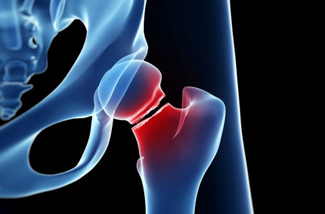
Le femur is the long thigh bone. Its upper end is composed of a ball-shaped head. The latter is connected to trochanter by a narrow zone called femoral neck. The trochanter (bulge) is a bony mass making the link between the femoral neck and the long part of the femur. The head of the femur articulates with the pelvic bone at a joint called hip joint.
La hip fracture is a broken femur bone, more specifically of theupper end of the femur. In other words, it corresponds to all the fractures that can affect the upper part of the femur (thigh bone). It can occur in three different places:
At the level of the neck of the femur, we then speak of cervical fracture;
At the level of the trochanter, we then speak of intertrochanteric fracture;
Either at the level of the head of the femur or under the large bulge (trochanter), we then speak of subtrochanteric fracture.
The causes of a hip fracture
At the people under 50, the fracture of the upper end of the femur usually occurs after severe trauma (road accident, fall from a high place, practice of a contact sport…).
At a Old person, hip fracture most often results from banal fall in 95% of cases. It overwhelmingly affects women.
THEosteoporosisis one of risk factorsmajor of a hip fracture. This disease causes the loss of bone mass in the neck of the femur and its fragility.
Alcohol abuse, prolonged use of corticosteroids or psychotropic drugs, smoking, thinness (because being overweight cushions the fall) and visual disturbances are also important risk factors. In addition, there are also pathologies occurring with age that promote falls, such as:
visual and auditory disturbances,
muscle weakness,
balance problems
other diseases that impair mobility (Parkinson's disease, stroke, etc.).
What are the symptoms of a fracture of the upper part of the femur?
Whatever the mode of onset, hip fractures lead in most cases to pain important and theimpossibilityto get up or lean on the leg.
The person eventually perceives a creak and feel a pain lively, localized at the level of the hip, in the groin crease or in the buttock. It can't walk, neither stand nor move the leg (functional impotence). Any support on the leg concerned is impossible. Palpation and mobilization of the thigh will cause pain. Sometimes the pain seems to be coming from the knee rather than the hip. This sensation is due to the fact that the knee and the hip have certain nerve pathways in common. This pain is called radiated pain.
La leg affected may seem shorter (shortened) and timespositioned (At external rotation). In other words, the patient will turn their foot outward. It is a reflex attitude, in order to avoid a painful posture. The thigh is deformed upwards.
If a large amount of blood leaks from the fracture, the person may feel dizziness or if feel weak. The area may swell (oedematous), and a purplish bruise may appear on the skin. This case is a surgical emergency.
How is the diagnosis made?
Le diagnostic is oftenposed on clinical examination. In general, the pains, the functional impotence and the deformity of the thigh allow the doctor to establish a diagnosis. In addition, in the event of a hip fracture, palpation and mobilization of the thigh cause pain.
Le initial radiological assessment includes a AP and lateral hip X-rays, a x-ray of the lower limb in internal rotation of 10˚ as well as a x-ray of the femoral neck in surgical profile. It is systematically associated with a pelvic x-ray face in search of a pelvic fracture associated.
X-rays will display le line of femur fracture. It will determine the type of fracture line (transverse, oblique, spiral, etc.), location of the fracture line (cervical, intertrochanteric and subtrochanteric), angulation (in degrees) and displacement bone fragments (in millimetres).
If the symptoms are present and the X-ray does not reveal a fracture line in the femur, it will be necessary to make a IRM (Magnetic resonance imaging) in order to refine the diagnosis and rule out any doubts (small fractures or fissures). The computed tomography (CT) is sometimes used, but is less accurate in detecting minor hip fractures.
How to treat a hip fracture?
In general, a hip fracture requires a interventionsurgical. It's about a surgical emergency. Ideally, this should be done within 24 hours of diagnosis to prevent the patient's general condition from deteriorating. The intervention can, however, be postponed in certain cases. The need to stop a contraindicated treatment during surgery such as certain oral anticoagulants is an example.
At a young person, the purpose of the processing is to consolidate the fracture as well as preserve vitality full femoral head. While at a Old person, his goal is to restore independence the most quickly as possible, in order to avoid the complications linked to prolonged bed rest and the deterioration of his state of health.
Some non-displaced fractures can however be managed without surgery, subject to strict bed rest.
It exists several types of surgical techniques the choice of which will depend on certain factors such as:
type of fracture;
shift ;
the age of the patient;
general state of health;
the importance of osteoporosis;
the patient's activity (profession, practiced sport, hobbies, etc.).
La hip prosthesis is especially recommended for elderly patients. This technique limits the duration of bed rest and allows the patient to recover more quickly.
The screws, pins, nails and plates on the other hand, are indicated for young patients. This with the aim of preserving the head of the femur and avoiding the installation of a hip prosthesis.
The different types of interventions to treat a broken hip
The hip prosthesis
La hip prosthesis involves replacing the head of the femur. When the roof of the hip joint (acetabulum) turns out to be in poor condition, the surgeon may have to replace it with a synthetic cup.
The prosthesis makes it possible to recover more quickly. It is indicated in particular after the age of 70 when it is essential to limit the duration of bed rest and therefore the appearance of complications.
The intervention varies according to the state of the capsule: a set of fabrics in the form of a sleeve which encloses the joint.
Before replacing the hip joint, doctors remove the broken pieces of bone.
If a partial hip prosthesis is needed, surgeons use a prosthesis designed to fit in the socket of the pelvic bone joint. The prosthesis has a solid stem that fits in the center of the femur. It is intended for elderly people who walk very little, and therefore put very little stress on the hip joint.
A total hip prosthesis is sometimes necessary, especially in the event of a femoral neck fracture which can disrupt the blood supply to the hip. It allows you to find a better function. It is increasingly common in older active people. During the placement of a total hip prosthesis, the femoral head and the surface of the corresponding cavity are replaced.
Screws, pins, nails and plates are preferred before age 70.
Femoral neck fractures can be repaired by inserting metal pins through the neck and into the head of the femur.
Intertrochanteric fractures can be repaired by attaching a sliding compression screw and a metal side plate to hold the bone fragments in their normal position while the fracture heals. It takes at least 6 months before people feel as comfortable and strong as before the incident and can walk as before.
How is rehabilitation going?
Whatever treatment is chosen, rehabilitation must be quicklyput implemented after the intervention.
The day after the operation, the patient is encouraged to take a few steps, with the help of canes or crutches, under the close supervision of a physiotherapist. You have to walk, but without forcing.
Possible complications after the procedure
General complications
They are favored by hospitalization and prolonged bed rest.
Phlebitis : A blood clot forms in the veins of the leg.
Pulmonary embolism : the clot can pass through the veins and reach the lungs.
Confusion mental.
Aggravation of an already existing disease (aggravation of diabetes, heart problem, etc.).
Bedsores (wound of the skin related to its prolonged compression).
Orthopedic complications
Several situations can arise.
Displacement of a fracture after osteosynthesis, often linked to osteoporosis.
Necrosis (destruction) of the femoral head. It is often the result of a displaced fracture of the femoral neck.
Infection bone or joint.
Pseudarthrosis that is to say the non-union of the fracture.
Complications related to the prosthesis
La most common complication of a hip replacement is the dislocation. The femoral part comes out of the cavity of the acetabular part. This situation generally occurs shortly after the intervention, following an accidental movement.
My name is Arotoky and I am studying human medicine. Web editor for more than 5 years, I currently focus on the theme of health and well-being. I have the strong conviction that with my valuable articles, I can help many people to relieve their ailments and feel better. My goal is to share my health knowledge with the general public through web writing.

")
")
")

")


